Dog Skeletal Skull Anatomy Poster 18 X 24 Etsy
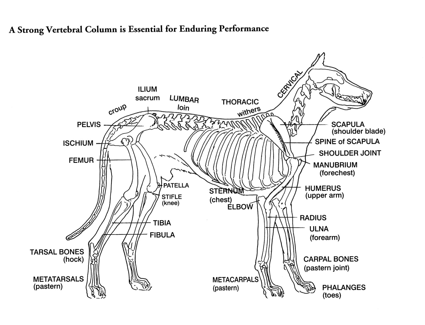
A dog's skeleton is formed so the dog can run fast, hunt and chase. For example, a dog's shoulder blades are not tightly connected to its skeleton, so the dog has potential for greater motion and flexibility. The dog skeleton has an average of 319 bones. Where Is the Skeleton Located in Dogs?

Skeletal abnormality in dogs and cats
Dog - Muscles Dog - Thorax/Abdomen/Pelvis Animal - Anatomy atlas: Cardiovascular system Veterinary anatomy - Animal: ANATOMICAL PARTS Abdomen Abdominal aorta Abdominal mammary gland Abdominal mammary region Accessory carpal bone Acromion Adductor muscle Ala of ilium; Wing of ilium Ala of nose Anconeus muscle Antebrachial region Aortic arch
A Visual Guide to Dog Anatomy (Muscle, Organ & Skeletal Drawings) All Things Dogs
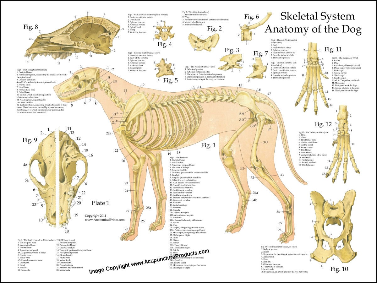
The dog skeleton is the bony part of dogs made for the support and protection of internal organs. Bones are connected through joints and muscles move the bones to produce the normal dog movements. In this article we will cover: Bone types and parts of the dog skeleton The dog skull Dog cranium The spine The Trunk The Forelimb The Hindlimb
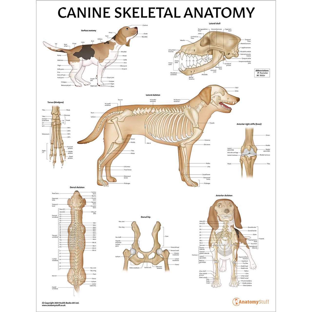
Canine Skeletal Anatomy Laminated Chart Dog Skeleton Poster
An overview of the anatomy of the canine skeleton.Follow on twitter @ https://twitter.com/PerkyVetInstagram: Perkydvm
Helen King on Structure Evaluation Susan Garrett's Dog Training Blog
25/04/2023 31/12/2021 by Sonnet Poddar The dog skeleton anatomy consists of bones, cartilages, and ligaments. You will find two different parts of the dog skeleton - axial and appendicular. Here, I will show you all the bones from the axial and appendicular skeleton with their special osteological features.
Dog Skeletal Anatomy
Speaking of skeletons, a dog has 320 bones in their body (depending on the length of their tail) and around 700 muscles. Muscles attach to bones via tendons. Depending on the breed of dog, they will have different types of muscle fibers. You've probably heard about slow and fast twitch muscle fibers before.

Скелет собаки Dog Skeletal Anatomy Dog Anatomy, Animal Anatomy, Anatomy Study, Anatomy
Figure 5-14 Detailed skeletal anatomy of the sacrum from a caudolateral view , sacrum and caudal 1 or Cd1 vertebra from a lateral view , Cd4 vertebra from a cranial view , and Cd6 vertebra from a dorsal view . (From Evans HE: Miller's anatomy of the dog, ed 4, Philadelphia, 2013, WB Saunders.)

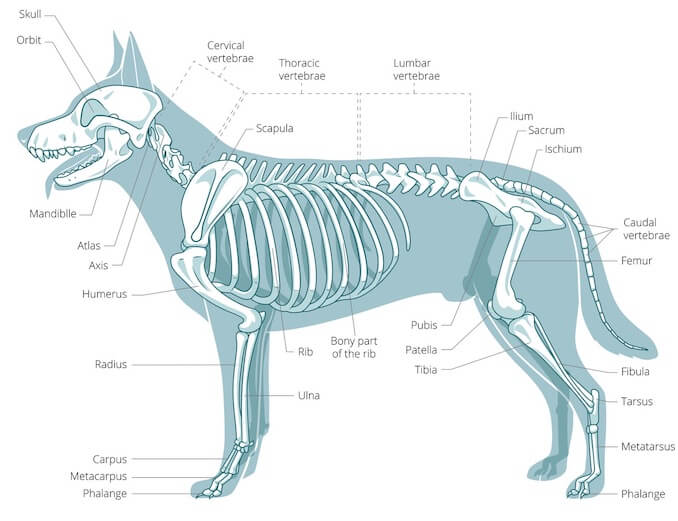
FileDog anatomy lateral skeleton view.jpg
The anatomy of a dog includes its skeletal structure, reproductive system, the internal organs, and its external appearance. The following paragraphs explain all these aspects in brief, along with diagrams, which will help you understand them better. External Anatomy Dogs, like all mammals, have eyes, a nose, a forehead, and ears.

PetMassage Chart 3 Skeleton of the Dog · PetMassage™ Training and Research Institute
Components of the Musculoskeletal System in Dogs By Stephen B. Adams , DVM, DACVS, Department of Veterinary Clinical Sciences, College of Veterinary Medicine, Purdue University Reviewed/Revised Mar 2018 | Modified Oct 2022 Bones provide rigid structure to the body and shield internal organs from damage.

Canine Anatomical Skeleton Bosley Dog (Large)
Buy Dog Skeleton Anatomy on ebay. Money Back Guarantee!
Canine Skeleton Poster Clinical Charts and Supplies
Dog anatomy comprises the anatomical studies of the visible parts of the body of a domestic dog.Details of structures vary tremendously from breed to breed, more than in any other animal species, wild or domesticated, as dogs are highly variable in height and weight. The smallest known adult dog was a Yorkshire Terrier that stood only 6.3 cm (2.5 in) at the shoulder, 9.5 cm (3.7 in) in length.

Dog Anatomy
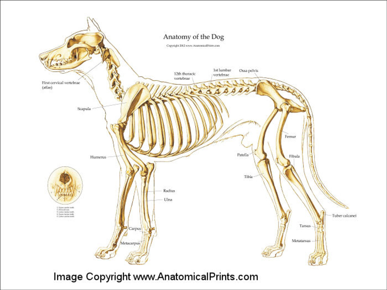
Here are presented scientific illustrations of the canine skeleton, with the main dog's bones and its structures displayed from different anatomical standard views (cranial, caudal, lateral, medial, dorsal, palmar..). Some of the different canine joints are labeled.

Vintage 1935 Dog Veterinary Print Skeleton Of Dog Anatomy Of Dog Canine Skeleton Dog Bones Book
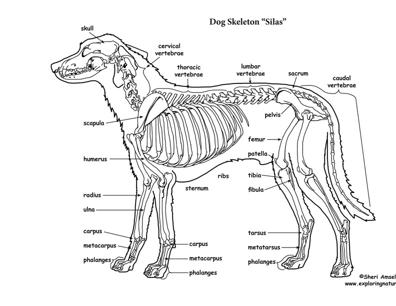
January 3, 2024 < http://www.exploringnature.org/db/view/Dog-Skeletal-Anatomy > Dog Skeletal Anatomy

Dog skeleton 101 Dog Anatomy Bones Animal Hackers
The cat has a small coronoid fossa medial to the radial fossa that accommodates the coronoid process of the ulna during elbow joint flexion.; The cat has a supracondylar foramen near the medial condyle allowing the passage of the median nerve and brachial blood vessels.; There is an intermediate tubercle between the greater and lesser tubercles in the horse's intertubercular groove.

Anatomy of dog skeleton with labeled inner bone scheme vector illustration Dog skeleton
The Anatomage Dog is the first highly detailed dog anatomy atlas that comprehensively features internal organs, including vascular systems and muscular-skeletal structures. Originating from real dog data, the Anatomage Dog exhibits the highest level of anatomical accuracy. All of its volumetric 3D and individual structures are segmented, users.

Anatomy Of Dog Spine Canis Lupus German Shepherd Skeleton Dog sketch, Dog anatomy, Dog skeleton
• Splanchnic skeleton, which in the dog and cat consists only of the os penis found within the soft tissues of the penis. • Each part of the skeleton consists of many bones, each of which plays an important part in the function of the skeletal system. • Bones are covered in 'lumps, bumps and holes'.